Rudraksha X-Ray Report Software: Storing, Labelling and Sharing Bead Images
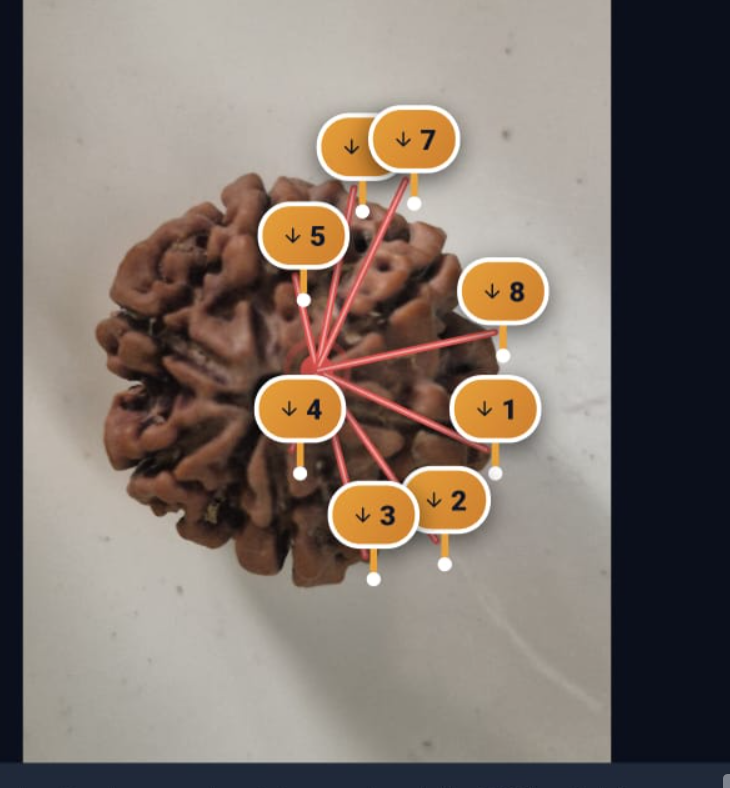
Deploying specialized rudraksha x-ray report software is the definitive step forward for modern gemological laboratories. In an industry where rare 1 to 21 Mukhi specimens are routinely fabricated by gluing separate segments or carving external furrows into low-Mukhi beads, digital radiography (X-Ray) provides the only indisputable forensic proof of biological authenticity.
However, capturing an X-ray scan is only half the battle. Testing laboratories must securely store high-resolution images, systematically label locular chambers, co-register radiographs with white-light photography, and share diagnostic proof with customers via instant cloud QR verification. Purpose-engineered rudraksha lab software solves this entire workflow seamlessly.
How Rudraksha X-Ray Report Software Transforms Locule Diagnostics
Unlike generic dental software or basic image viewers that treat X-rays as isolated pictures, LabAuthentiq integrates radiographic files directly into the specimen's master diagnostic record:
✓ 1. Dual-Spectrum Co-Registration
The software pairs the macro white-light photograph (showing external flutes, color, and surface spikes) side-by-side with the digital radiograph. Technicians can toggle between spectra to confirm that every external Mukhi corresponds to an internal seed chamber.
✓ 2. Automated Locule Chamber Counting & Soundness Scoring
Technicians inspect the radiopaque seeds inside each compartment. Using the interactive workstation, they annotate natural furrows, hairline fissures, or glued joints. The software computes a real-time Structural Soundness score (Grade A Akhanda down to Grade C Khandita) and generates official certificate remarks automatically.
✓ 3. Edge Convolution for Fake Carve & Glue Detection
Built-in 3×3 Sobel mathematical gradient filters highlight unnatural adhesive glue lines and synthetic saw-cuts invisible to the naked eye. If a counterfeiter joined two 4-Mukhi halves to create an 8-Mukhi bead, the radiograph edge analysis exposes the glue seam instantly.
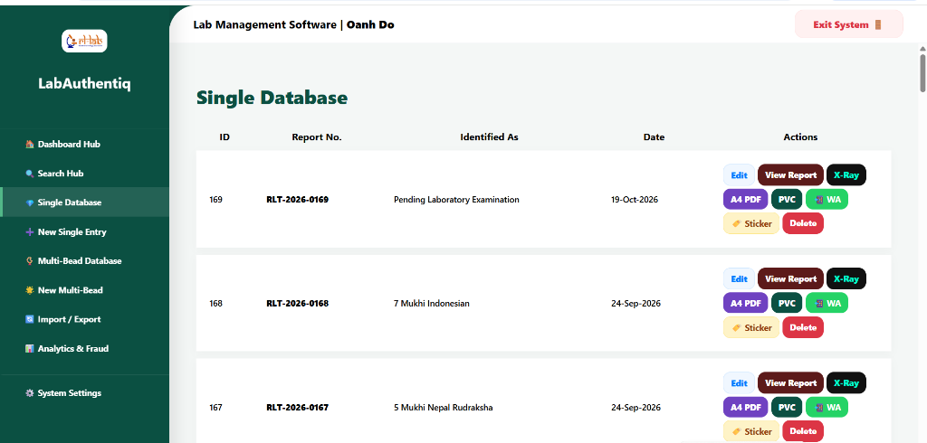
Cloud Storage & Inode Architecture: Preserving 10,000+ Specimen Archives
Digital radiographs are high-resolution files (typically 2 MB to 8 MB each). Storing thousands of scans on a local desktop computer creates serious vulnerabilities: hard drive crashes, accidental deletion, and zero remote accessibility.
LabAuthentiq SaaS bundles 1 Year of High-Speed NVMe Cloud Hosting with 150,000 Inodes. This robust infrastructure allows laboratories to archive over 10,000 complete testing dossiers—including original raw radiographs, processed contrast scans, macro photography, and generated Smart PVC cards—with automated daily offsite backups.
Automated Multi-Bead Mala & Combination Dossier Generation
Testing an individual bead is simple; testing a 54 or 108-bead Siddh Mala or Kantha used to require 3 to 4 hours of tedious manual formatting in Microsoft Word. Technicians had to crop 108 photos, align 108 X-ray scans, and pray the margins didn't break.
With LabAuthentiq's specialized rudraksha lab software, mala generation is 100% automated. The software compiles every bead's macro photo, X-ray scan, individual weight, and locule tally into a stunning, printable A4 multi-page booklet or A3 master spread in under 30 seconds.
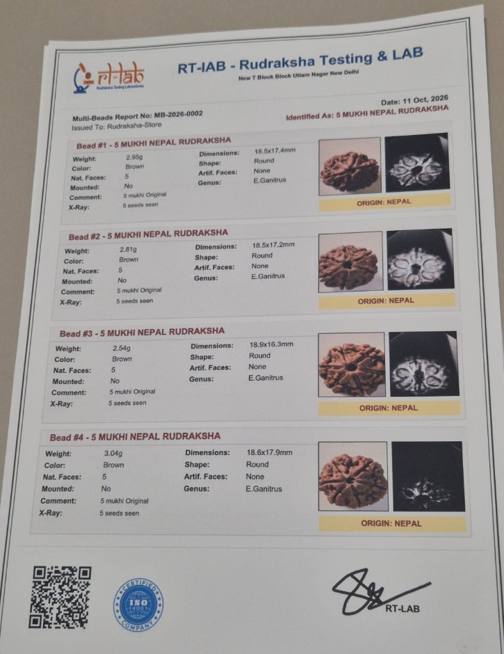 Multi-Bead Mala Technical Dossier Output
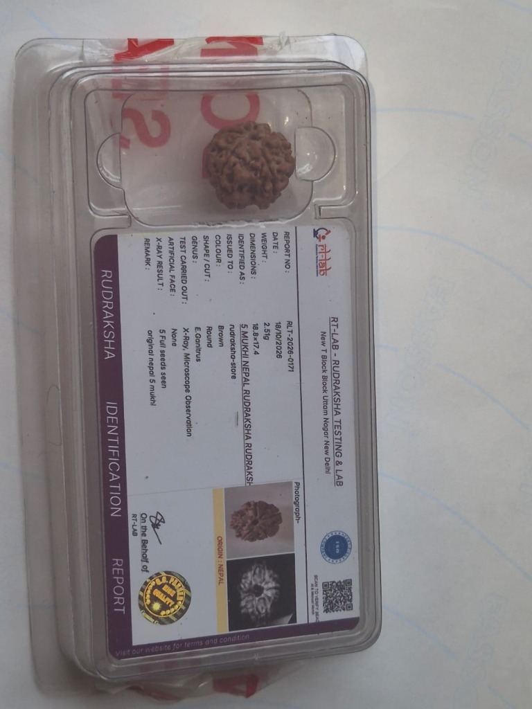 Instant Smart PVC Card with Locule Count Badge
Recommended Technical Guides & Whitepapers
Frequently Asked Questions (FAQ)
Why is specialized software required for Rudraksha X-Ray radiography? ↓
Standard image viewers cannot co-register cross-sectional radiographs with external macro photography, nor can they calculate locule counts or apply edge filters. LabAuthentiq Rudraksha X-Ray report software pairs both imaging modalities into a permanent certificate record.
How does the software store large X-Ray files securely? ↓
LabAuthentiq includes 1 Year of High-Speed NVMe Cloud Hosting with 150,000 Inodes, allowing laboratories to store over 10,000 complete specimen dossiers—including high-resolution radiographs and Smart PVC print queues—with automated daily backups.
Can clients view the original X-Ray on their phones? ↓
Yes. When clients scan the certificate's QR code, the software renders an interactive high-resolution radiograph viewer directly in their smartphone browser, allowing pinch-to-zoom locule verification without needing any app install.
Does the software support multi-bead mala X-Ray grids? ↓
Yes. LabAuthentiq includes an automated multi-bead mala engine that arranges 27, 54, or 108 individual bead photos and X-ray radiograph pairs into clean, printable A4 multi-page and A3 master spread dossiers in seconds.
Streamline Your Lab's X-Ray Workflow
Upgrade to LabAuthentiq SaaS today. Experience dual-spectrum co-registration, automated locule counting, and 150,000 Inode cloud archiving.